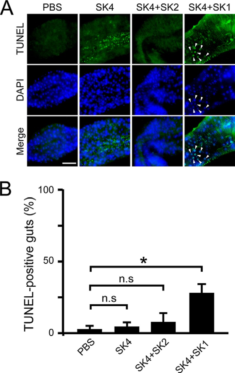
FIGURE 7.

TUNEL assays in the midguts of gnotobiotic flies that mono- or co-ingested strains SK1–4. A, TUNEL staining of apoptotic cells in the fly midgut after 18 days of continuous ingestion. Images show the copper cell region of the midgut. Arrowheads, representative TUNEL-positive cells. Green, TUNEL-positive cells; blue, DAPI nuclear stain; scale bar, 50 μm. B, the percentage of guts showing apoptosis-positive signals was calculated. The values shown are means ± S.E. (error bars) (n = 3). p values were calculated by one-way ANOVA followed by the Bonferroni post hoc test. *, p < 0.05; n.s, not significant.
