FIGURE 3.
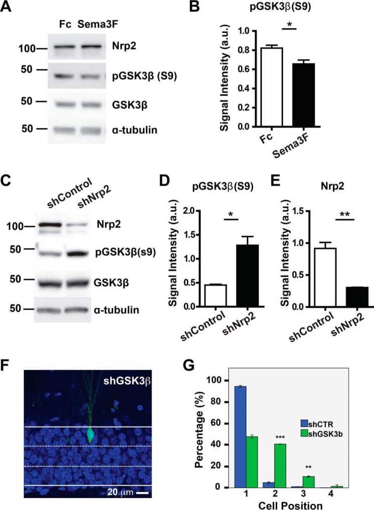
Stimulation of NRP2 by Sema3F activates GSK3β and consequently affects cell positioning. A, representative Western blots showing serine phosphorylation of GSK3β (Ser-9) (pGSK3β (S9)) upon stimulation of NRP2 by Sema3F-Fc in primary hippocampal neurons, with anti-GSK3β, anti-NRP2, and anti-tubulin antibodies as loading controls. B, graph shows the quantification for signal intensity of Western blot bands for phosphorylated GSK3β. a.u., arbitrary units. C, representative Western blots show that serine phosphorylation of GSK3β is increased in NRP2 knockdown cells. Knockdown efficiency of shNRP2 was shown with anti-NRP2 antibody, while anti-GSK3β and anti-tubulin antibodies were used as loading controls. D and E, graphs show the quantification for signal intensity of Western blot bands for phosphorylated GSK3β and NRP2. F, representative image of shGSK3β-expressing newborn neurons in the DG at 28 DPI (scale bar, 20 μm). G, quantification of cell positioning of adult-born neurons expressing shCTR (blue bars) or shGSK3β (green bars). *, p < 0.05,**, p < 0.01, ***, p <0.001, t test.

