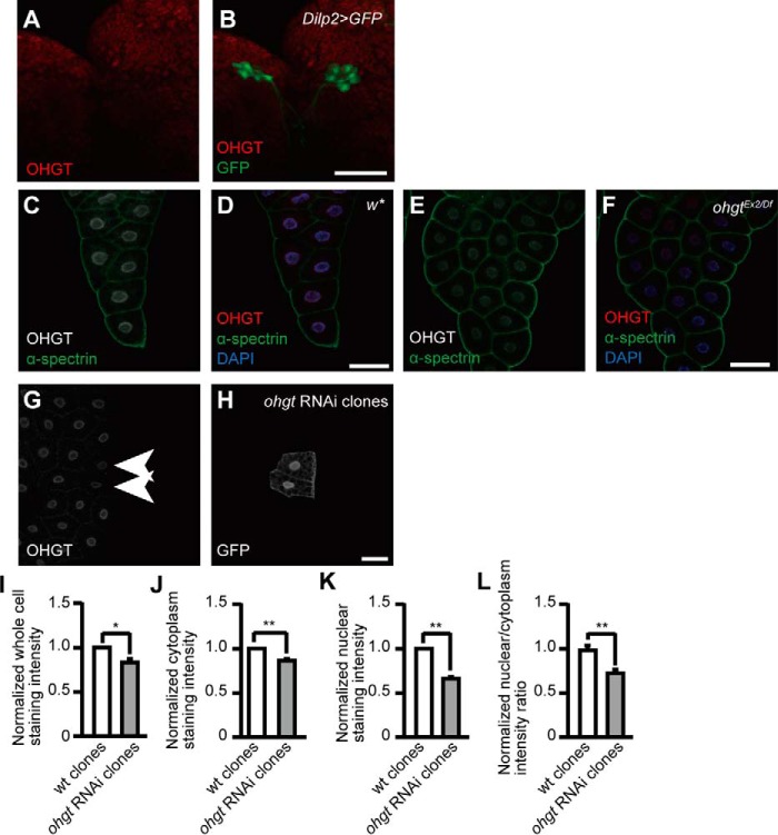
FIGURE 4.
OHGT is expressed in the nucleus of fat body cells. A and B, co-immunostaining of OHGT and GFP in the IPCs of dilp2>GFP third instar larva. C and D, co-immunostaining of OHGT and plasma membrane marker α-spectrin in the fat body of w* early third instar larva (72 h AED). E and F, co-immunostaining of OHGT and α-spectrin in the fat body of ohgtEx2/Df early third instar larva (72h AED). G and H, co-immunostaining of OHGT and GFP after clonal ohgt RNAi induction in fat body cells. Genotype, y,hs-flp/+; Dcr-2/+; act-FRT-CD2-FRT-Gal4, UAS-GFP/UAS-ohgtRNAiv40486. The scale bars in images represent 50 μm. I–L, normalized staining intensities in whole cells, the cytoplasm, and the nucleus as well as the nuclear to cytoplasm ratio of wild type clones (n = 34) and ohgt RNAi induced clones (n = 17). Error bars indicate S.E. * indicates p < 0.05, and ** indicates p < 0.01. p values were calculated by Mann-Whitney U test.

