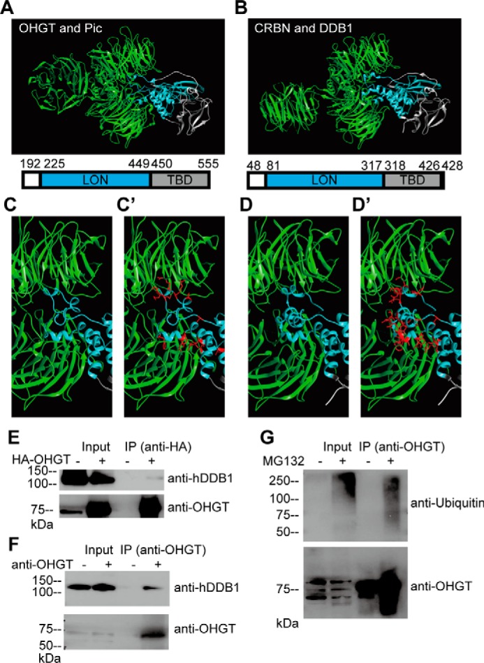
FIGURE 7.

OHGT interacts with PIC, the Drosophila homolog of human DDB1. A, 3D model for OHGT-PIC complex was created using the SWISS-MODEL service and then aligned to respective positions in Protein Data Bank code 4TZ4. Amino acid residues and domains depicted in the structures are shown. B, the crystal structure of the human CRBN-DDB1 complex (Protein Data Bank code 4TZ4). C and D, a close-up view of the OHGT-PIC/CRBN-DDB1 interaction interfaces. C′ and D′, amino acid side chains of OHGT/CRBN that point toward the PIC/DDB1 interface are shown in red. E, S2 cells were transfected with HA-OHGT expression vector, and protein complexes containing HA-OHGT were immunoprecipitated using HA tag-specific antibody. Total lysates (Input) and immunoprecipitates (IP) were subjected to Western blotting with antibodies to the indicated proteins (for detecting OHGT, guinea pig OHGT-specific antibody was used). F, protein complexes containing endogenous OHGT were immunoprecipitated using rabbit OHGT-specific antibody. Total lysates (Input) and immunoprecipitates (IP) were subjected to Western blotting with antibodies to the indicated proteins (for detecting OHGT, guinea pig OHGT-specific antibody was used). G, S2 cells were treated with MG132, and endogenous OHGT was immunoprecipitated using rabbit OHGT-specific antibody. Total lysates (Input) and immunoprecipitates (IP) were subjected to Western blotting with antibodies to the indicated proteins (for detecting OHGT, guinea pig OHGT-specific antibody was used).
