
FIGURE 1.
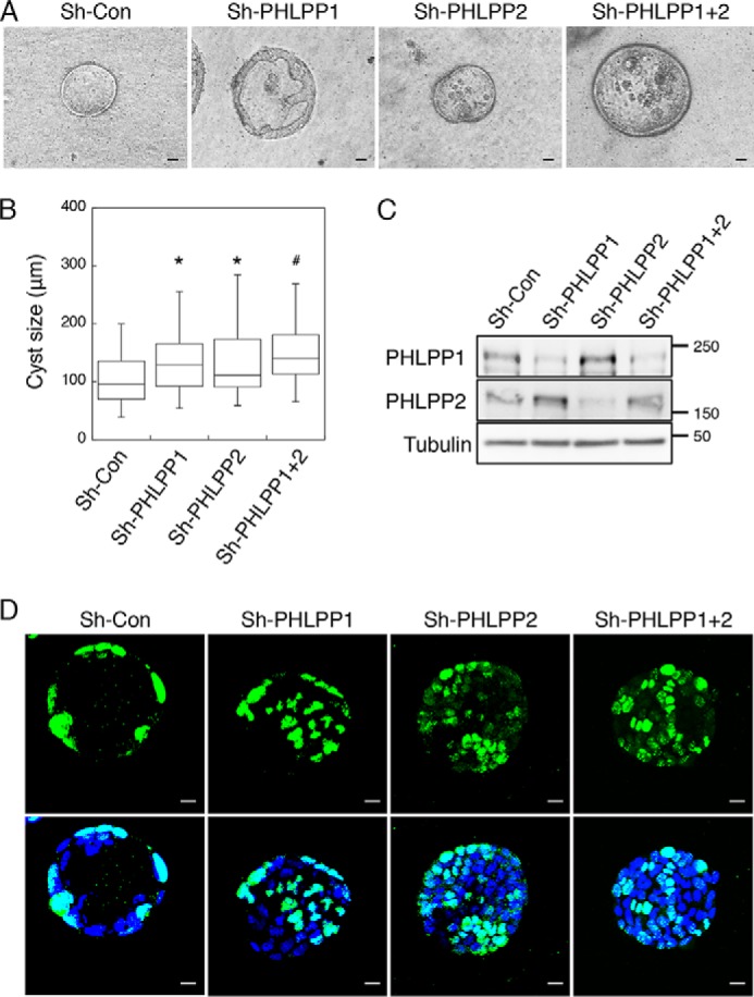
Knockdown of PHLPP alters morphogenesis of Caco2 cells grown in three dimensions. Caco2 cells infected with shRNA lentivirus targeting PHLPP1, PHLPP2, or both PHLPP isoforms were seeded in 3D matrix as a single cell suspension and allowed to grow for 14 days. A, the representative phase-contrast images of the cyst-like structure formed by control and PHLPP knockdown cells. The images were obtained using a Nikon TE2000 inverted microscope with 10× objective. Scale bar, 20 μm. B, the longest diameter of cysts formed by the control and PHLPP knockdown cells was measured using the Nikon Elements AR software. The size distributions of 50 randomly chosen cysts were analyzed for each group of cells and are shown in the box-whisker plot. The average cyst sizes for the following cells are (means ± S.E., in μm): sh-Con, 105.5 ± 5.7; sh-PHLPP1, 140.2 ± 8.5; sh-PHLPP2, 132.4 ± 7.8; and sh-PHLPP1+2, 146.8 ± 6.5 (n = 50, * indicates p < 0.01 and # indicates p < 0.001 by Student's t test when compared with sh-Con cells). C, the expression of PHLPP1 and PHLPP2 in the control and PHLPP knockdown cells as determined using Western blotting. D, the control and PHLPP knockdown cells grown in three dimensions were fixed and stained with the Ki67 antibody (green) and DAPI (blue). Confocal images of stained cells were obtained using an Olympus FluoView FV1000 confocal laser-scanning microscope. Scale bar, 20 μm. Note that the cysts formed by PHLPP knockdown cells show a lack of fully polarized structure and an increased number of Ki67-positive cells.