FIGURE 5.

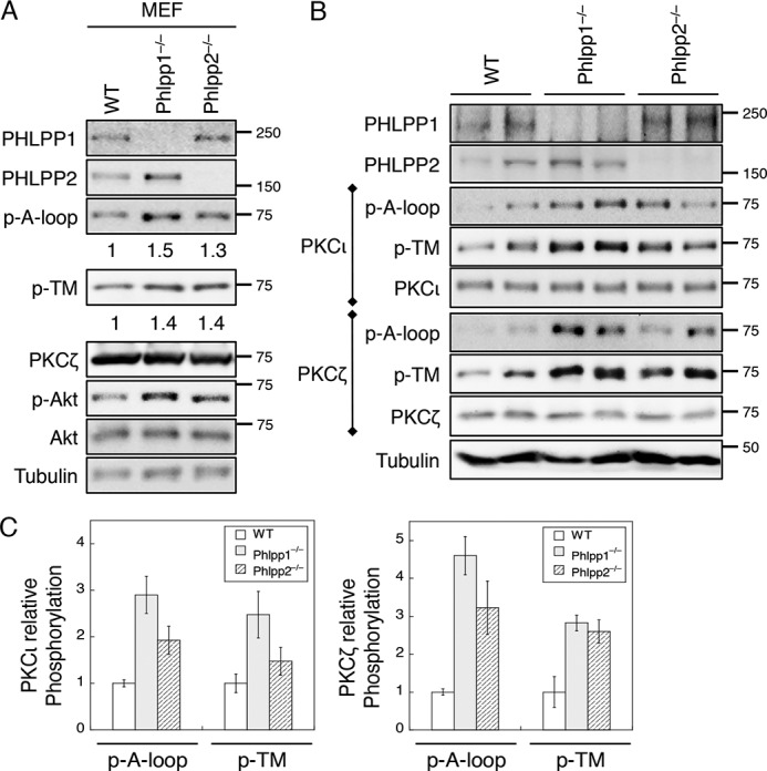
The phosphorylation of aPKCs is increased in PHLPP knock-out MEF cells and mouse tissues. A, cell lysates prepared from WT, PHLPP1−/−, and PHLPP2−/− MEF cells were analyzed for the phosphorylation (p) of PKCζ at the A-loop and TM sites using the phospho-specific antibodies. The relative phosphorylation was quantified by normalizing the amount of phosphorylation as detected by the phospho-specific antibody to that of total protein and shown below the phosphoblots. B, fresh colon tissues were isolated from WT, PHLPP1−/−, and PHLPP2−/− mice. Two mice from each genotype were analyzed. Protein extracts were prepared and first immunoprecipitated with the PKCι or PKCζ antibody. The phosphorylation status of PKCι and PKCζ was determined using the phospho-specific antibodies. C, Western blots as shown in B were quantified by normalizing the amount of phosphorylation as detected by the phospho-specific antibody to that of total protein. Graphs show the average results of two mice. Data shown in the graphs represent the means ± S.D.
