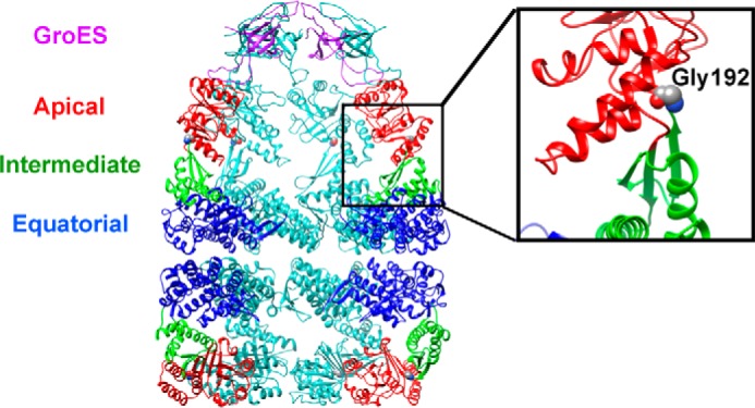
FIGURE 1.

Structural model of GroE, showing the location of Gly-192 in the GroEL subunit. The figure on the left is a cutaway image of the GroEL14·ADP7·GroES7 complex, derived from Protein Data Bank (PDB) file 1AON (36). Selected GroEL and GroES subunits have been removed to showcase the characteristic central cavity of GroEL14. The GroEL subunits shown in the forefront of the figure have been colored to highlight the domain architecture; the apical domain is in red, the intermediate domain is in green, and the equatorial domain is in blue. The panel on the right is an expanded view of the hinge II region of the GroEL subunit, with Gly-192 represented as space-filling forms. Images were drawn using UCSF Chimera (37).
