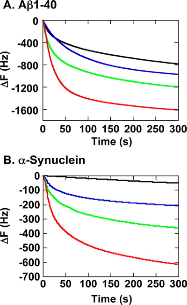
FIGURE 5.

QCM binding analyses of GroEL WT and three G192X mutants to immobilized fibrillogenic polypeptides. A, the binding of GroEL to immobilized Aβ(1–40) peptide. B, the binding of GroEL to immobilized αSyn-His6. In each panel, the black trace indicates the binding of GroEL WT, the blue trace indicates the binding of GroEL G192N, the green trace indicates the binding of GroEL G192I, and the red trace indicates the binding of GroEL G192W.
