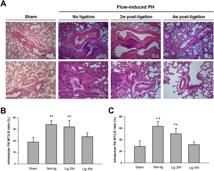
Figure 7.
Representative lung sections of animals with and without flow-induced pulmonary hypertension (A). Morphometric analysis of the medial thickness of the intralobular (ϕ 500–1000 μm, (B) and intraacinar (ϕ <250 μm, C) pulmonary artery. Thickness of medial layer is expressed as wall thickness to lumen radius (WT/LR) ratio (%). Thickness of medial layer was significantly reduced 4 weeks (lig 4w) after ligation of aortocaval fistula. *P < 0.05 compared with sham and †P < 0.05 compared with lig 4w. Magnifications are 100× for the upper panel and 200× for the lower panel. Analysis was performed in 5–6 different lung sections in each group, and the medical thickness was measured in at least 1 intralobular and 2 intraacinar pulmonary artery in each tissue section using the ImageJ software. Data were analyzed by one-way ANOVA and are shown as mean ± SD.

