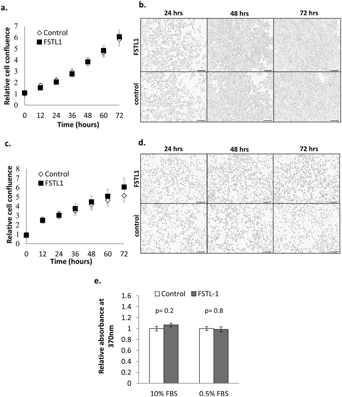
Figure 9. FSTL-1 does not regulate β cell growth and proliferation.
INS-1 β-cells were plated at a density of 1.5 × 10⁴ cells/well in a 96 well plate. Post synchronisation, the cells were either provided with untreated medium or medium supplemented with 100 ng/ml rFSTL-1 in either 10% FBS (a,b) or 0.5% FBS (c,d) and cultured for a further 72 hrs. Cell growth was monitored every 12 hrs during this period using the IncuCyteZOOM live cell imaging system. In (a,c) graphs shows mean cell confluence relative to control +/− SEM, n = 30 from 5 independent experiments, while representative images taken at the indicated timepoints are shown in (b,d). BrdU incorporation was measured for the last 24 hrs of the 72 hr culture, and the graph in (e) shows mean absorbance relative to the control +/− SEM. Statistical significance was measured using Student’s t-test (unpaired, two-tailed) and p-values are indicated in the graph.

