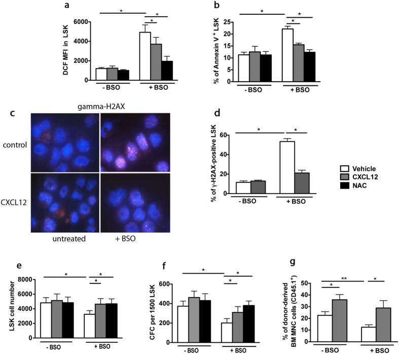
Figure 5. CXCR4/CXCL12 axis prevents BSO-induced oxidative stress in HSCs.
(a) DCF MFIs of sorted WT LSK cells cultured for 48 hr with vehicle or BSO, in the absence or presence of 100 ng/mL CXCL12 or 2 mM NAC. Mean ± SEM, n = 6. (b) Annexin-V staining of LSK cells. Mean ± SEM, n = 3. (c) γ-H2AX-staining of LSK cells cultured for 48 hr in the absence (vehicle) or presence of BSO and CXCL12. Representative pictures are shown. (d) Percentages of γ-H2AX-positive LSK cells. More than 100 cells were counted per group in 3 independent experiments. (e) Effect of BSO-treatment on LSK proliferation. Mean ± SEM, n = 4. (f) Colony numbers (CFC) from LSK cells. Mean ± SEM of colonies from 5 independent experiments performed in duplicate. (g) CXCL12 rescues compromised hematopoietic long-term reconstitution with BSO-treated LSK cells. Before transplantation into lethally irradiated WT CD45.2+ recipients, CD45.1+ LSK cells were treated with BSO for 48 hr in the absence or presence of CXC12. Mean ± SEM of percentages of CD45.1+ donor-derived cells 6 months after transplantation from the pool (10 mice) independent experiments.

