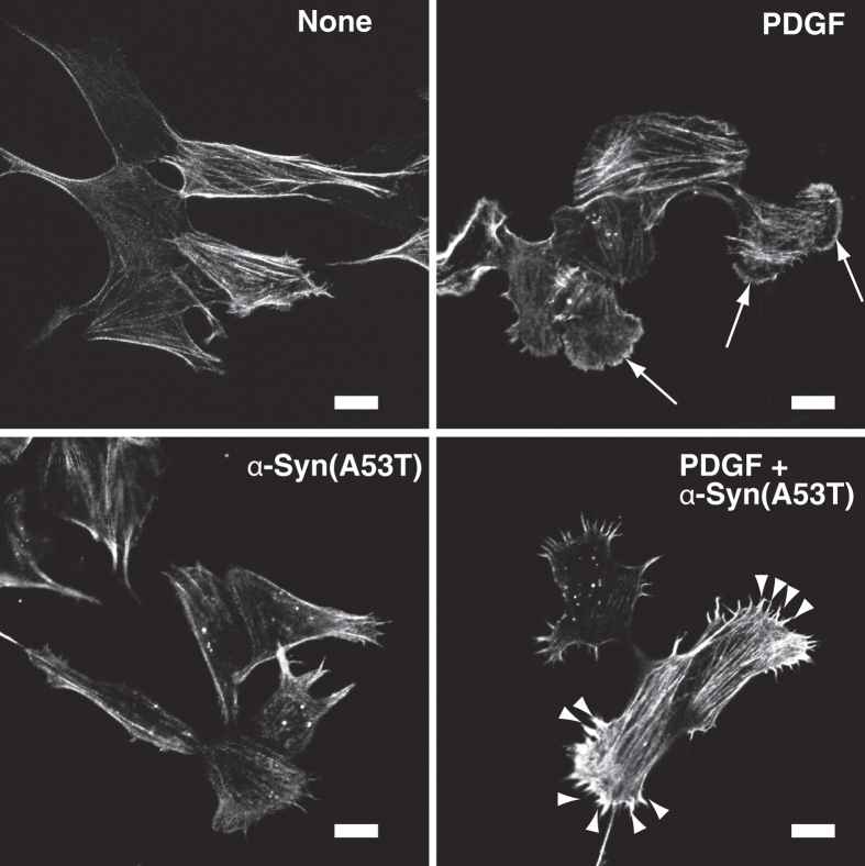
Figure 7. α-Syn(A53T)-caused abnormal actin cytoskeleton rearrangement.
SH-SY5Y cells, which had been cultured in the absence of serum for 18 hr without or with 1 μM α-Syn(A53T), were stimulated for 8 min without (none) or with 20 ng/ml PDGF. After fixation, actin filaments were stained by rhodamine-phalloidin. The results are the representative of 3 independent experiments. Arrows show PDGF-induced lamellipodia formation. Note that PDGF causes filopodia (see arrow heads) instead of lamellipodia in α-Syn(A53T)–treated cells. Bars, 10 μm.

