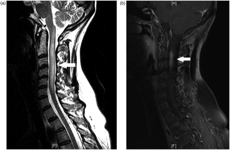
FIGURE 2.
(a) Sagittal view of T2 image of MRI of the cervical spine showing a longitudinal lesion, extending from C1 through the inferior level of C6 with expansion of the cord. (b) Sagittal view of T1 post-contrast MRI of the cervical spine showing enhancement of the cervical cord predominantly at C2–C3.

