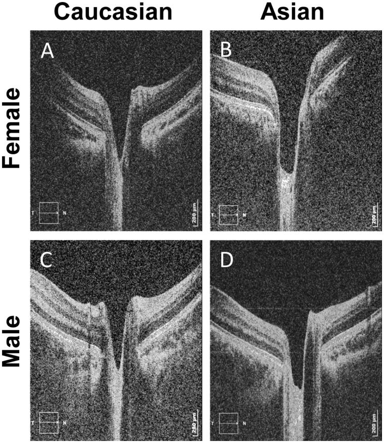
FIGURE 1.
Tomograms of the optic nerve head. Right eyes of (A) a 31-year-old Caucasian female, (B) a 34-year-old Indian female, (C) a 35-year-old Caucasian male and (D) a 27-year-old Indian female participant. Refer to the scale of the scan in the right bottom corner of each tomogram where the horizontal axis is 106 pixels/mm and the vertical axis is 513 pixels/mm.

