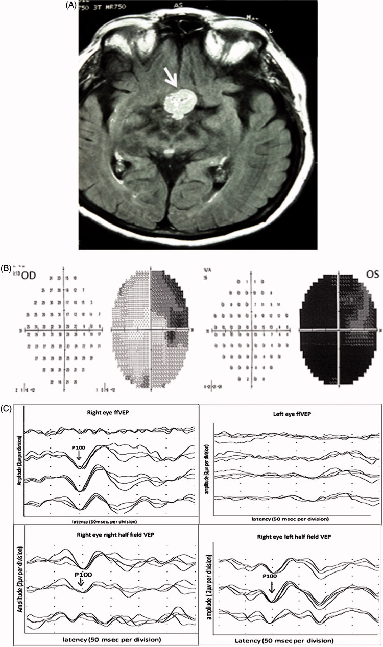
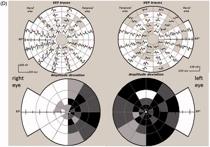
FIGURE 2.
(A) Axial T1-weighted MRI with contrast showing a large mass (white arrow) occupying pituitary fossa and extending into the suprasellar cistern and right cavernous sinus consistent with a pituitary tumour. (B) Humphrey 24-2 visual fields showing total visual field loss in the left eye and upper temporal scotoma in the right eye. (C) Full-field PVEP of both eyes showing no consistent response in the left eye and normal amplitude and latency in the right eye. The half-field PVEP of the right eye shows smaller amplitude in the right half with normal latency in both half-fields. (D) mfVEP showing total reduction of amplitude in the left eye and temporal hemianopia in the right eye.


