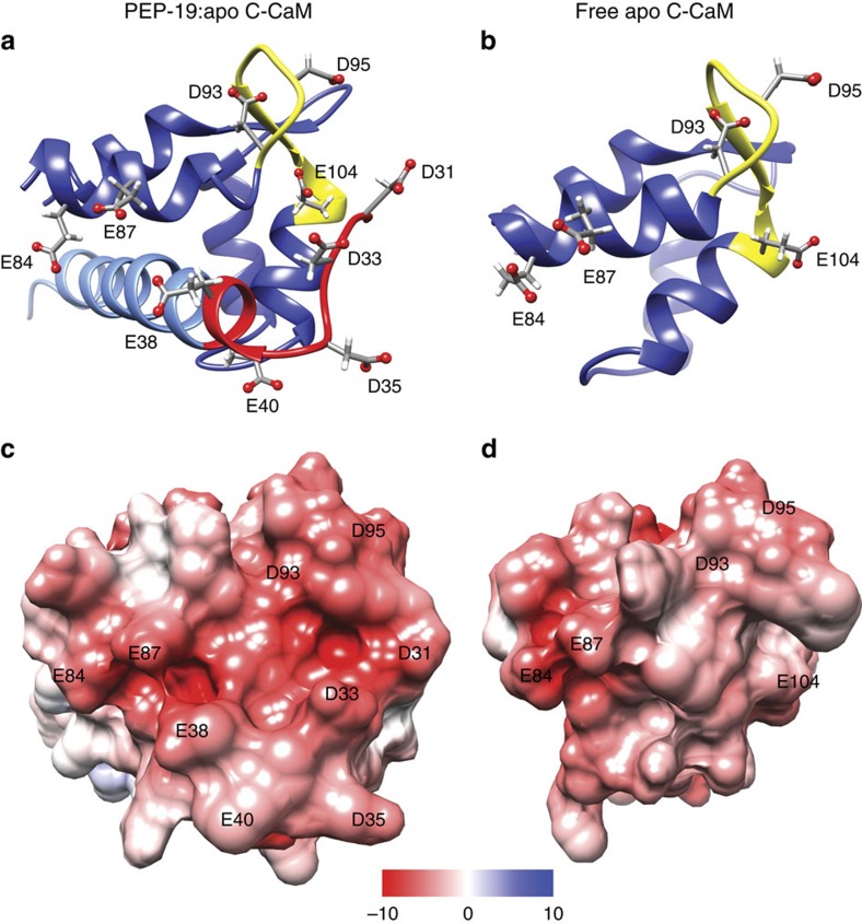
Figure 5. The acidic sequence of PEP-19 greatly increases the negative ESP near Ca2+ binding site III of apo C-CaM.
(a,b) Ribbon diagrams for the PEP-19:apo C-CaM complex and free C-CaM, respectively. Dark blue is C-CaM, yellow is Ca2+ binding loop III, red and light blue are the acidic sequence and core IQ motif in PEP-19, respectively. (c,d) Solvent excluded surfaces that are coloured based on ESP.

