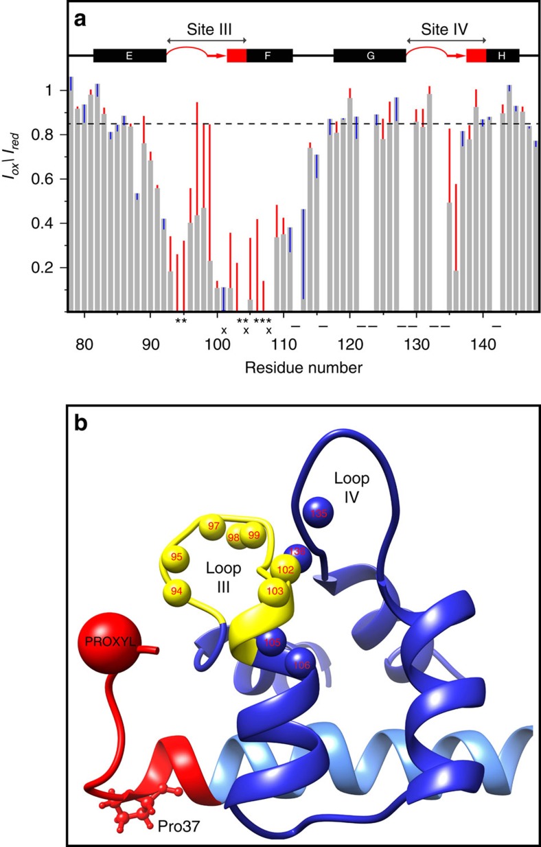
Figure 6. Mutation of Pro37 to Gly in PEP-19 increases the distance between the acidic sequence and Ca2+ binding site III in apo C-CaM.
Asp31 in PEP-19 and PEP(P37G) was mutated to Cys and spin labelled for PRE analysis as described in the methods. The grey bars in (a) the normalized amide Iox/Ired intensity ratios derived from 1H, 15N HSQC spectrum of apo C-CaM when bound to PEP-19(SL). The red and blue vertical lines indicate an increase or decrease, respectively, in Iox/Ired due to mutation of Pro37 to Gly. The Iox/Ired could not be calculated for residues indicated by (−) due to spectral overlap. Amide cross peaks for residue indicated by (*) are line broadened beyond detection when C-CaM is bound to PEP-19(SL), while those indicated by (x) are line broadened beyond detection when bound to PEP(P37G)SL. (b) The location of amides (small balls) for residues in apo C-CaM that show the greatest increase in distance from the PROXYL probe with an increase in Iox/Ired of greater than 0.2 due to mutation of Pro37 to Gly. Calcium binding loop III is shown in yellow.

