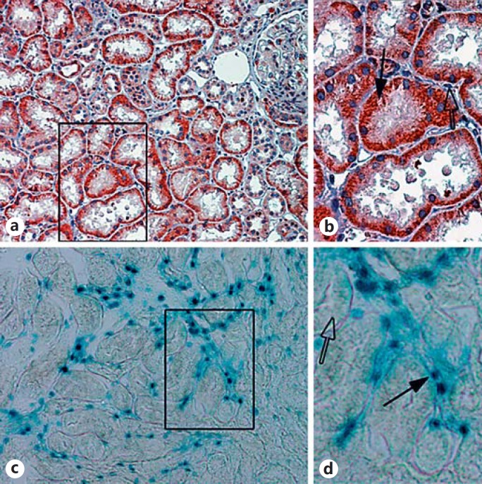
Fig. 3.
Shh is produced by renal tubules but acts on interstitial fibroblasts. a Immunohistochemistry for Shh after ischemia-reperfusion injury is shown with red indicating positive staining. Note the strong staining within injured tubules. b Inset showing an enlarged image of the boxed area in a. Shh staining is specific for renal tubules (black arrow) but not interstitial cells (open arrow). c Mice with the LacZ reporter under control of the Gli promoter show that only interstitial cells exhibit increased β-galactosidase activity (denoted as blue staining). d Inset showing an enlarged image of the boxed area in c. Gli reporter activity is specific for interstitial cells (black arrow) but is not present in tubular cells (open arrow).

