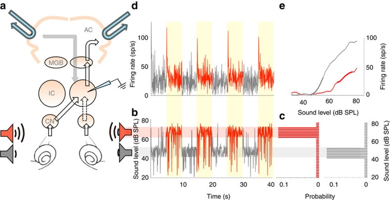
Figure 1. Auditory midbrain responses to switches between sound-intensity environments.
(a) Recording set-up. Electrode in IC. IC receives input from ears via contralateral cochlear nucleus (CN), and, indirectly, ipsilateral CN (dotted line). Auditory cortex (AC) receives from IC via medial geniculate body (MGB) and also gives bilateral feedback. Cooling loops (blue). (b) Section of broadband noise sound stimulus, quiet (grey) and loud (red) environments. (c) Level distribution per environment type. (d) Firing rate over time of single neuron to stimulus. (e) Firing rate versus stimulus intensity of neuron per environment. Superimposed thick lines represent high-probability regions of c.

