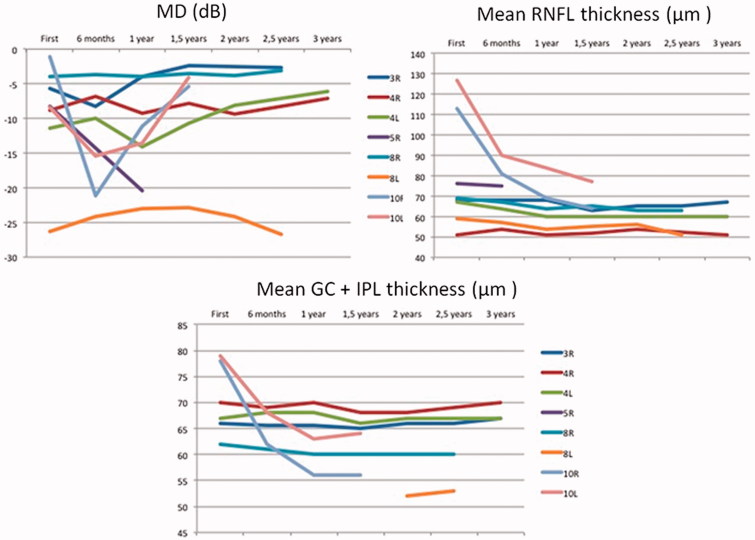
FIGURE 1.
Visual field and RNFL and GC thickness changes during the follow-up period of three years. Case 5 and 10 showed significant worsening of their visual fields due to recurrent tumoral growth. RNFL and GC thickness remained stable save for case 10 who initially showed papilledema that disappeared after surgery.

