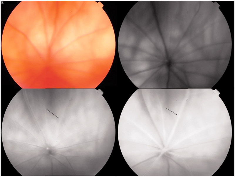
FIGURE 3.
Typical fluoroscein fundus angiography (FFA) performed on BCCAO rats before surgery. Upper left: fundus image; upper right: blue reflectance (red-free) image. The retina was observed after fluorescein sodium was injected into the caudal vein. Arterial filling appeared at 5.7 s (lower left) and complete engorgement at 6.7 s (lower right).

