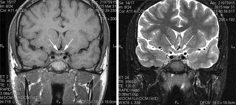
Figure 2.

Noncontrast MRI at 2 weeks after initial visual loss. T1-weighted thin coronal section through chiasm (right) demonstrates hypointense central regions bilaterally (arrows). T2-weighted thin coronal section same plane (left) shows hyperintense central regions bilaterally (arrows).
