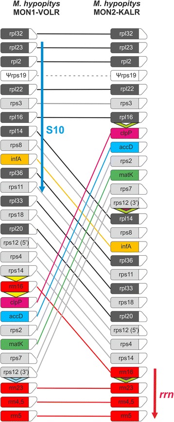
Fig. 3.

Schematic gene order comparison maps between the plastid genomes of M. hypopitys MON1-VOLR and M. hypopitys MON2-KALR. The linear representation of the circular mapping genomes using rpl32 genes as the starting point does not reflect the actual gene size or spacing between the coding regions. Boxes represent protein-encoding and rRNA genes with the direction of transcription shown by the chamfered top corner. Triangles in M. hypopitys maps indicate repeat sequences. The S10 and rrn operons are shown by arrows. Note that comparison of the structures of plastid genomes of M. hypopitys and Nicotiana tabacum is shown in Additional file 4: Figure S4
