
Fig. 2.
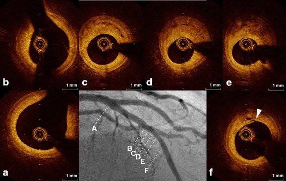
OCT image of the middle left anterior desceinding artery lesion revealed a coronary plaque with homogeneous high signal intensity (Fig. 2b, c, d), and clearly visualized an intraplaque neovascular microchannel (NVMC) network arising from the distal portion (arrowhead, Fig. 2f) to the tightest lesion (Fig. 2d). The NVMC network spread further proximally, and was distributed within the intra-medial plaque, not connected to the adventitial vasa vasorum (Fig. 2c). There were no obstructions at other sites of the LAD (Fig. 2a, b)