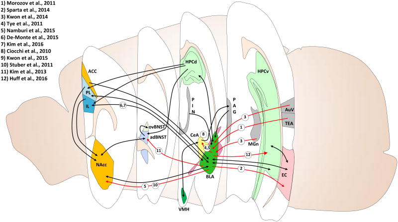
Figure 1. Neural circuits involves in fear and anxiety-related behaviors in rodents.
Optogenetic, electrophysiological, and pharmacogenetic techniques have elucidated many specific circuitries underlying rodent fear and anxiety-related behaviors. Cross sectional views taken from different anterior-posterior positions within the rodent brain are marked with relevant brain regions and their distal projections. Projections highlighted in red are discussed in the present review; these highlighted circuits account for only a portion of identified circuitries, some of which are labeled with black arrows. ACC, anterior cingulate cortex; adBNST, anterodorsal nucleus of the BNST; AuV, secondary auditory cortex; BLA, basolateral amygdala; CeA, central amygdala; EC, entorhinal cortex; HPCd, dorsal hippocampus; HPCv, ventral hippocampus; IL, infralimbic division of the mPFC; MGn, medial geniculate nucleus; NAcc, nucleus accumbens; ov, oval nucleus of the BNST; PAG, periaqueductal gray; PIN, intralaminar thalamic nuclei; PL, prelimbic division of the mPFC; TEA, temporal association cortex; VMH, ventromedial hypothalamus.

