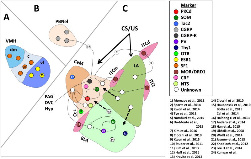
Figure 2. Microcircuits and specific neuronal populations in the amygdala, ventromedial hypothalamus (VMH) and parabrachial nucleus (PBN) involved in fear and anxiety-related behaviors.
A) Microcircuits and cell populations in the ventromedial hypothalamus. B) PBN projections to the CEA. C) Amygdala microcircuits and subnuclei. Known microcircuits discussed in the present review are noted; dashed black arrows denote projections between amygdala subnuclei. Forked lines indicate glutamatergic projections whereas crossed lines indicate GABAergic projections. BLA, basolateral amygdala; c, central division of the ventromedial hypothalamus; CEAm, medial subdivision of the central amygdala; CEAl, lateral subdivision of the central amygdala; CGRP, calcitonin gene-related peptide; CGRP-R, calcitonin gene-related peptide receptor; dm, dorsal medial division of the ventromedial hypothalamus; DVC, dorsal vagal complex; ESR1, estrogen receptor; Hyp, hypothalamus; ITC, intercalated cell nuclei; ITCd, dorsal intercalated cell nuclei; ITCl, lateral intercalated cell nuclei; ITCm, medial intercalated cell nuclei; MOR, mu opioid receptor; OT, oxytocin; PAG, periaqueductal gray; PBNel, external lateral subdivision of the PBN; PKCd, protein kinase C delta; PV, parvalbumin; SF1, steroidogenic factor 1; SOM, somatostatin; Tac2, tachykinin 2; vl, ventrolateral division of the ventromedial hypothalamus; VMH, ventromedial hypothalamus.

