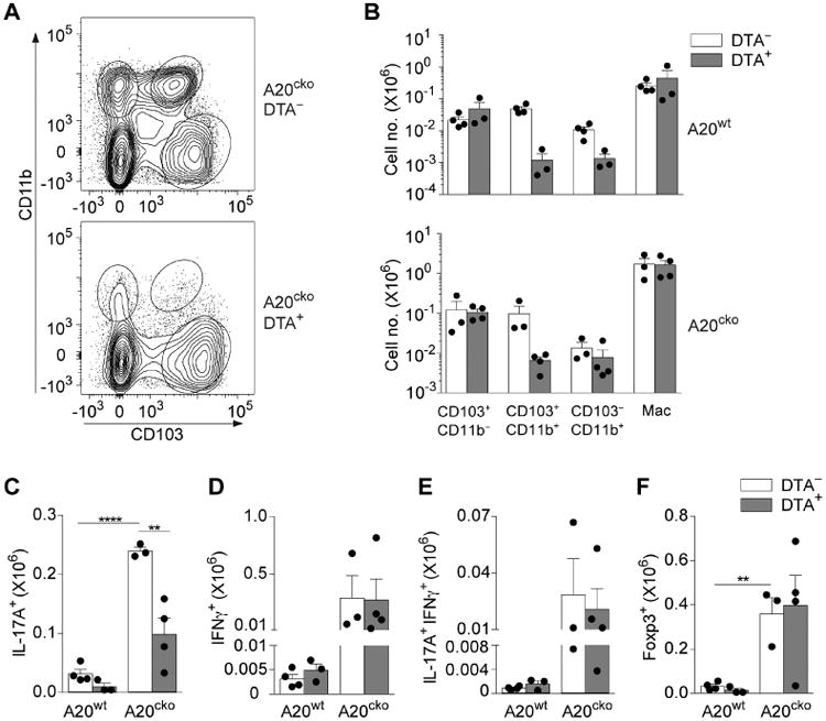
Figure 7. In small intestine inflammation CD103+CD11b+ DCs and CD103−CD11b+ DCs expand inflammatory Th17 cells.

(a) Representative flow plot of SI-LP DCs in co-housed, littermate A20cko-DTA− mice and A20cko-DTA+ mice at 9-12 weeks of age. (b) Absolute number of each SI-LP DC subset in A20wt or A20cko mice, either DTA− or DTA+. (c-f) Absolute number of SI-LP CD4 T cells, either IL-17+ (c), IFNγ+ (d), IL-17+IFNγ+ (e) or Foxp3+ CD4 T cells (f) in co-housed A20wt or A20cko mice, additionally either DTA− or DTA+. Results were combined from 3 independent experiments including at least one mouse of each genotype. Each dot represents a mouse. Error bars represent mean ± SEM. *, P < 0.05, **, P < 0.01, ****, P < 0.0001, (unpaired student's t-test).
