
Figure 6. UV Survival and Rates of CPD Removal of Yeast Carrying Different Rad4 Variants.
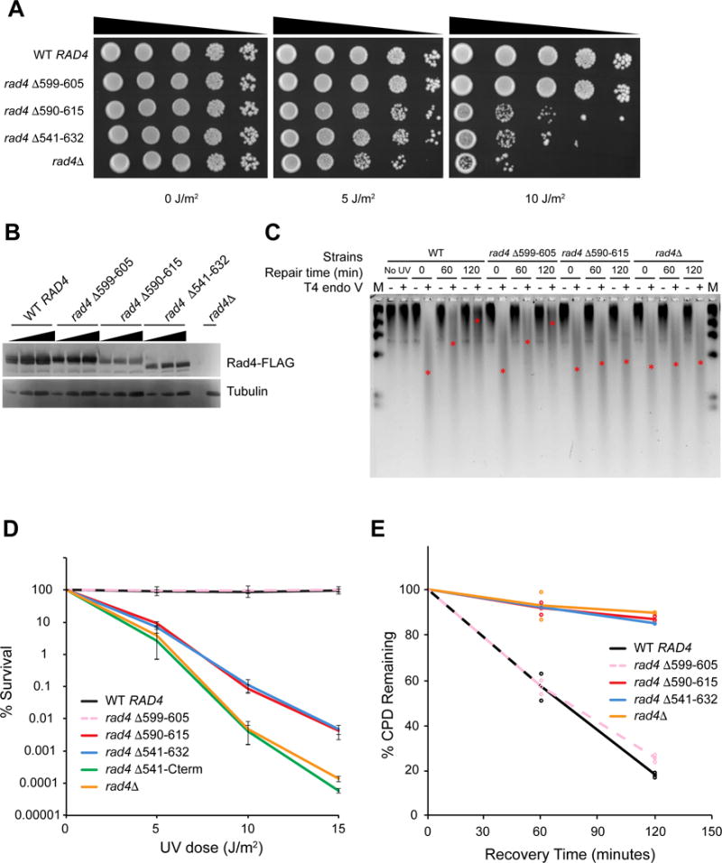
(A) Serial dilutions of yeast cells (BY4742) expressing different 3×FLAG-tagged Rad4 variants on YPD plates, 72 hours after UV irradiation.
(B) Expression levels of 3×FLAG-tagged Rad4 variants detected with anti-FLAG antibody.
(C) Genomic DNA of yeast cells after UV irradiation and recovery digested with T4 endo V, separated on alkaline agarose gel, and detected with SYBR Gold. Approximate positions of the ensemble average size of DNA in each lane are denoted with red asterisks (*). DNA marker (M, λ DNA-HindIII) was loaded in the left- and right-most lanes.
(D) Quantitative UV-survival of yeast cells (BY4741) expressing different untagged Rad4 variants. WT RAD4 – black, rad4 Δ599–605 (Δβ-hairpin3) – pink dashed, rad4 Δ590–615 – red, rad4 Δ541–632 (ΔBHD3) – blue, rad4 Δ541-cterm – green, rad4Δ – orange.
(E) Quantitative rates of CPD removal of yeast cells (BY4741) expressing different untagged Rad4 variants, determined by T4 endo V digestion. Color scheme same as in Figure 6D. See also Figure S7.