
Fig. 2. Fimbrial biogenesis models.
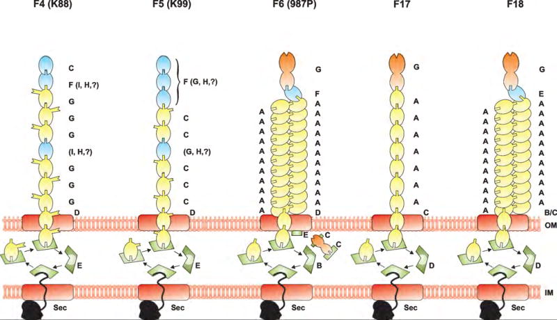
The fimbriae consist all of the polymeric assembly of a major subunit (yellow) and of one or more minor subunits (blue), one of them being a tip adhesin (orange) for some fimbriae. For the K88 and K99 fimbriae, the major subunit is the adhesin. Usher proteins (red) locate in the outer membrane and channel the fimbrial subunits to the bacterial surface. All the fimbrial export systems use one periplasmic chaperone (green) for all the subunits, with the exception of the F6 fimbriae that have three chaperones, two being dedicated to two different fimbrial subunits. All the fimbrial proteins cross the inner membrane by using the general secretion (Sec) pathway (black), with the exception of fimbriae specific regulators that remain in the cytoplasm (not shown).