Figure 3.
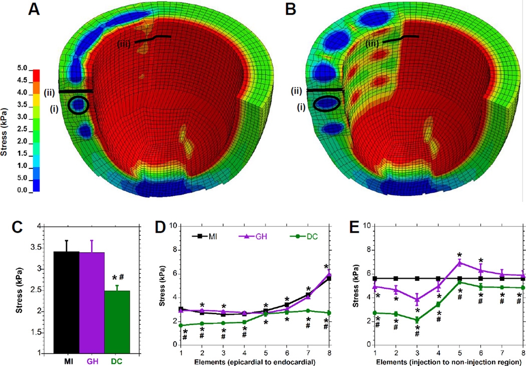
Finite Element Analysis of Hydrogel Injection. A-B, End-diastolic myofiber stress distribution for an LV with either (A) GH hydrogel injection or (B) DC hydrogel injection. Note that only a portion of the model is shown in order to visualize the distribution within the myocardium. C-E, Myofiber stress in elements adjacent to the material (C), or distributed along a transmural (D) or circumferential (E) path in the edge of the injection region. Corresponding regions are indicated for the material region (i) or transmural (ii) and circumferential (iii) paths. Data are presented as mean±SD with error bars for all points (some not visible due to low error); n≥6; *P < 0.05 relative to MI; #P < 0.05 relative to GH.

