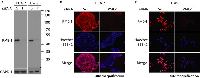
Figure 1.

Validation of the specificity of PME‐1 antibody in colorectal cancer cell lines. (A) Western blot image of protein lysates from HCA‐7 and CW‐2 cells transfected with scrambled (S) or PME‐1 (P) siRNA (for 72 h), and blotted with PME‐1 (B‐12) antibody. GAPDH was used as a protein loading control. Black lines denote the location of protein molecular weight marker bands. Immunofluorescence images of HCA‐7 (B) and CW‐2 (C) cells transfected with Scr or PME‐1 siRNA (for 72 h), and incubated with PME‐1 antibody and visualized with anti‐mouse‐Alexa‐594 secondary antibody (red). Hoechst 33342 shows nuclear staining (blue). PME‐1 and nuclear staining overlay is shown in merge (fuchsia). All images were taken at 40× magnification.
