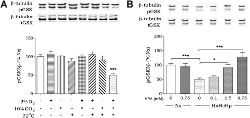
Figure 1.

Developing an in vitro model of in vivo haemorrhagic shock. (A) Huh7 cells were exposed to hypoxia, hypercapnia and/or hypothermia for 4 h as indicated and analysed for pGSK3β‐Ser9 levels and normalized to Nx. (B) Huh7 cells were exposed to normoxic conditions (37°C, 5% CO2; Nx), or combined hypoxia, hypercapnia and hypothermia (HxHcHp), treated with VPA (0.75 mM) as indicated, analysed for pGSK3β‐Ser9 levels and normalized to Nx. Data were quantified from at least triplicate experiments with technical triplicates (n > 9) ± SEM. Data were analysed using one‐way ANOVA and post hoc Tukey test.
