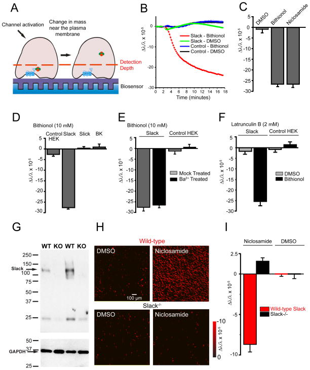
Figure 1. Stimulation of Slack channels alters mass distribution at the plasma membrane.
(A) Schematic diagram of Slack activation in a cell adherent to the biosensor. (B) Activation of Slack-expressing, but not untransfected, HEK cells with bithionol (10 μM) produced a sustained decreased in mass near the plasma membrane, n=32 wells/condition, p<0.001. (C) Changes in mass in Slack-expressing HEK after Slack activation with bithionol (10 μM) or niclosamide (500 nM) (n=16, p<0.001). (D) Stimulation of Slack, but not other Slo channel family members, with bithionol (10 μM) decreases mass near the plasma membrane, n=24, p<0.001. (E) Blocking ion flux through Slack channels with Ba2+ (1 mM) does not alter the signal upon channel activation, n=16. (F) Latrunculin B (2 μM) does not alter the signal upon Slack activation by bithionol (10 μM), n=12. (G) Slack protein expression is absent in brain synaptosomes isolated from Slack−/− mice (n=3/genotype). (H and I) Activation of Slack channels in WT, but not Slack−/−, murine cortical neurons decreases mass near the plasma membrane. (H) Representative images showing change in mass in cortical neurons with red representing decreases in mass and black representing no change in mass. (I) Quantification of (H), n=12, p<0.001. Error bars ±SEM.

