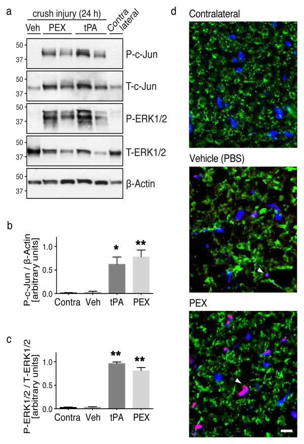
Figure 3. Activation of c-Jun in SCs by LRP1 ligands in vivo.
(a) Immunoblot analysis was performed to detect phosphorylated c-Jun (P-c-Jun) and activated ERK1/2 (P-ERK1/2) in crush-injured rat sciatic nerves injected with 2 μl of PEX (20 μM), tPA (12 μM), or vehicle. Uninjured contralateral nerve was studied as a control. Nerves were injected 24 h after crush-injury and harvested 15 min after injecting ligands. Equal amounts of nerve protein (20 μg) were loaded into each lane and subjected to SDS-PAGE. Immunoblot analysis was performed to detect β-actin and total ERK1/2 as loading controls. Each lane represents a different rat. (b) Quantification of phosphorylated c-Jun relative to β-actin by densitometry (n=4 rats/group; * denotes p < 0.05; ** denotes p < 0.01 compared to contralateral nerve). (c) Quantification of phosphorylated ERK1/2 relative to total ERK1/2 by densitometry (n=4 rats/group; ** denotes p < 0.01 compared to contralateral). (d) IF microscopy to detect phosphorylated-c-Jun (red). Nerves were injected with vehicle, or PEX, 24 h after crush injury. Nerves were collected 15 min later. Contralateral nerves were examined as uninjured controls. Transverse cryosections (10 μm) of sciatic nerve tissue, 1–3 mm distal to the injury site, were examined. The compact myelin protein, P0 (green), was stained to highlight SC architecture. Images are at 60x magnification (scale bar 10 μm). Note c-Jun immunoreactivity co-localized with DAPI-stained nuclei (blue) in SC crescents (pink). Images represent n=2/group.

