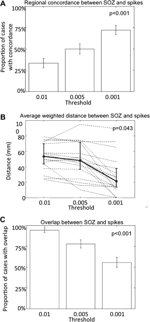
Fig. 4.

Relationship between spikes and the seizure onset zone. The most prominent spikes are detected close to but not always in the seizure onset zone. (A) The concordance between the region (defined as mesial temporal, inferior temporal, lateral temporal, frontal, parietal or occipital) containing the highest number of prominent spikes and the region comprising the seizure onset zone increases with the detection of only the most prominent spikes. Error bars indicate ±1 standard error of proportion (p < 0.001; one-tailed Fisher exact test). (B) The average distance between contacts containing spikes, weighted for the number of spikes detected by contact, and the seizure onset zone as a function of the prominence of detected spikes: more prominent spikes (detected with more stringent thresholds from 0.01 to 0.001) are closer to the seizure onset zone, as shown by the decreasing average distance. Dashed lines indicate individual cases and the bold line is the group mean; error bars indicate ±1 standard deviation. (p = 0.043; Kruskal–Wallis test). (C) The probability of overlap between the seizure onset zone and the contacts containing the prominent spikes decreases when only the most prominent spikes are detected. Error bars indicate ±1 standard error of proportion (p < 0.001; one-tailed Fisher exact test).
