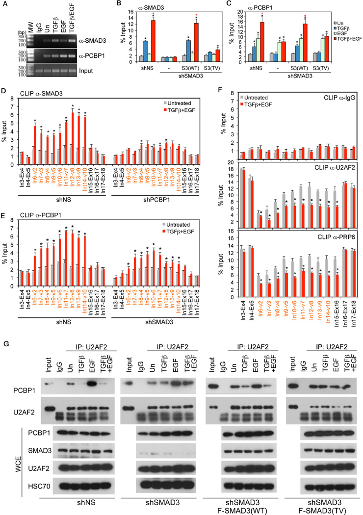
Figure 5. Formation of the SMAD3 and PCBP1 complex on variable exon regions of CD44 pre-mRNA precludes spliceosome assembly.
A: RIP assays of SMAD3 and PCBP1 binding to CD44 pre-mRNA in HeLa cells with growth factor treatment for 2 hr.
B-C: qRT-PCR analyses of RIP assay for SMAD3 (B) and PCBP1 (C) in HeLa cells.
D-E: qRT-PCR analyses of CLIP assay for SMAD3 (D) or PCBP1 (E) binding to intron-exon junctions (red denotes variable intron-exon junctions) of CD44 pre-mRNA in HeLa cells.
F: CLIP assays for control non-specific IgG, as well as U2AF2 or PRP6 binding to intron-exon junctions (red denotes variable intron-exon junctions) of CD44 pre-mRNA in HeLa cells.
G: IP-Western analyses of PCBP1 and U2AF2 following IP of U2AF2 from whole cell extracts (WCE) of HeLa cells expressing different vectors and treated with growth factors for 2 hr.
All bars are shown as mean ± SD (n=3). Statistical significance (p<0.02) is indicated for treated (2 hr) versus untreated samples (black *) and samples treated with TGF-β and EGF versus those with TGF-β alone (Red *).
See also Figure S5.

