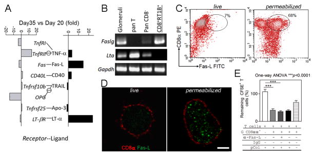
FIGURE 4.
CD8αα+RT1B/D+Ly49s6+ cells express intracellular Fas-L. (A) qPCR array for apoptosis pathway on glomerular mRNAs at early inflammation (day 20 post immunization) and T cell apoptosis peak (day 35) of immunized WKY rats; apoptosis-inducing receptors and ligands are shown separately and paired; expression of each gene is expressed as fold changes between day 20 and day 35; note that significant increases are seen in both Faslg and Ltα. (B) RT-PCR shows the detection of a high level of Faslg mRNA in the isolated glomeruli-infiltrating CD8αα+RT1B/D+Ly49s+ cells (CD8+RT1B+), but no Ltα expression was detected. (C) Two color flow cytometry on isolated glomeruli-infiltrating CD8αα+RT1B/D+Ly49s6+ cells for the detection of Fas-L; permeabilized cells showed a Fas-L+CD8α+ population (gated). (D) Immunofluorescence shows intracellular Fas-L (green) in a permeabilized CD8αα+RT1B/D+ cell (red) (right panel), but not in a live one (left panel). Bar =3 μm. (E) Killing assay shows that addition of Fas-L Ab (α-Fas-L) did not inhibit CD8αα+RT1B/D+Ly49s6+ cell-mediated killing of pCol(28–40) specific T cells. Three independent experiments were conducted with similar results. One-way ANOVA with Tukey post test was conducted. ***, p<0.001.

