FIGURE 5.
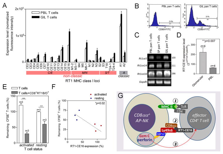
Activated autoreactive T cells with elevated level of RT1CE16 are more susceptible to the killing by CD8αα+RT1B/D+Ly49s6+ cells. (A) Summary of classic (RT1A) and non-classic (RT1C/E, M/N and S/T) MHC class I gene cluster expression pattern in PBMC (P) and glomeruli-infiltrating (GIL) T cells; expression level is normalized intensities in DNA microarray. n=4. (B) Flow cytometry shows detection of T cell activation marker CD69 in PBL pan T cells or glomeruli-infiltrating T cells as indicated. (C) Conventional RT-PCR detection of representative non-classic MHC I genes in PBL, splenic (SP), and glomeruli-infiltrating T cells. (D) Quantitative PCR detection of Rt1ce16 expression in glomeruli-infiltrating or PBL T cells. Relative expression levels to house-keeping gene Gapdh are shown. (E) Summary of susceptibility of T cells to CD8αα+RT1B/D+Ly49s6+ cell-mediated killing. Killing efficacy is expressed as % of remaining CFSE-labeled target T cells. T cells alone were used as control. (F) Correlation between RT1CE16 expression level in resting and activated pCol(28–40)-specific T cell lines and their susceptibilities to CD8αα+RT1B/D+Ly49s6+ cell-mediated killing; RT1CE16 expression levels were measured by qPCR and expressed as % of average for activated T cells; linear progression was calculated; each sample was duplicated. (G) Schematic diagram depicts three-step killing of autoreactive T cells by CD8αα+RT1B/D+Ly49s6+ cells (renamed to CD8αα+ AP-NK cell): Step 1, Coupling of CD8αα+ AP-NK cell with effector autoreactive T cell through its Ag presentation. Step 2, Binding of Ly49s/CD8αα on CD8αα+ AP-NK cell to RT1CE16 on the T cell. Step 3, DAP12 mediated release of granule cytotoxic proteins Gzm C and perforin from CD8αα+ AP-NK cell, which induce apoptosis in the target T cell.

