
FIGURE 6. Both DENV and soluble NS1 are released during blood feeding from infected mosquitoes.
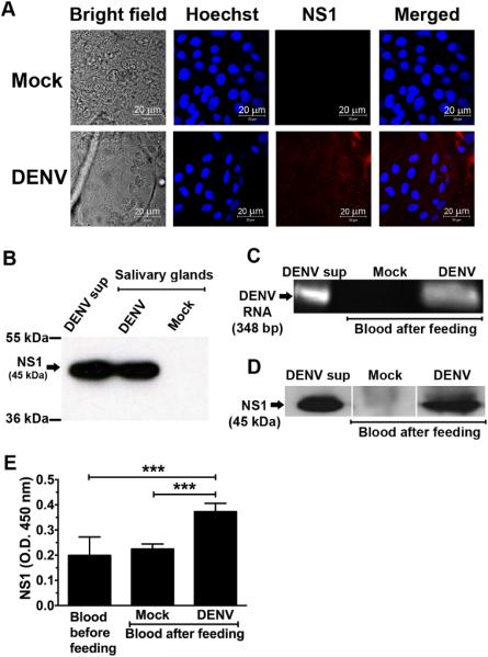
A. Representative bright field and immunofluorescence images of the salivary glands of mock- and DENV-infected mosquitoes. Aedes aegypti female mosquitoes were challenged orally with DENV by feeding them with an infectious blood meal and incubated for 14 days. Salivary glands of mosquitoes were harvested, smeared, and fixed on glass slides. NS1 was detected using a mouse anti-DENV NS1 mAb 1F11 (1 μg/ml) followed by Cy3-conjugated goat anti-mouse IgG. Nuclei were stained with Hoechst, a DNA-specific dye. Salivary glands harvested from uninfected mosquitoes served as a negative control. Analysis was performed by confocal microscopy. B. Expression of NS1 in the salivary glands of DENV-infected mosquitoes was analyzed by Western blotting. Fifty-fold concentrated supernatants from DENV-infected C6/36 cells (DENV sup) and the salivary glands harvested from uninfected mosquitoes (Mock) served as positive and negative controls, respectively. C-D. At 14 days after initial challenge with DENV, a total of 600 to 1200 mosquitoes were fed with a second non-infectious blood meal, the presence of DENV and NS1 in the remaining blood were determined by RT-PCR (C), Western blotting with DENV NS1 specific mAb 1F11 (D) and NS1-ELISA (E), respectively. Error bars indicate SD corresponding to three independent experiments. Asterisks denote the means that are statistically different among indicated groups (*** P < 0.001).