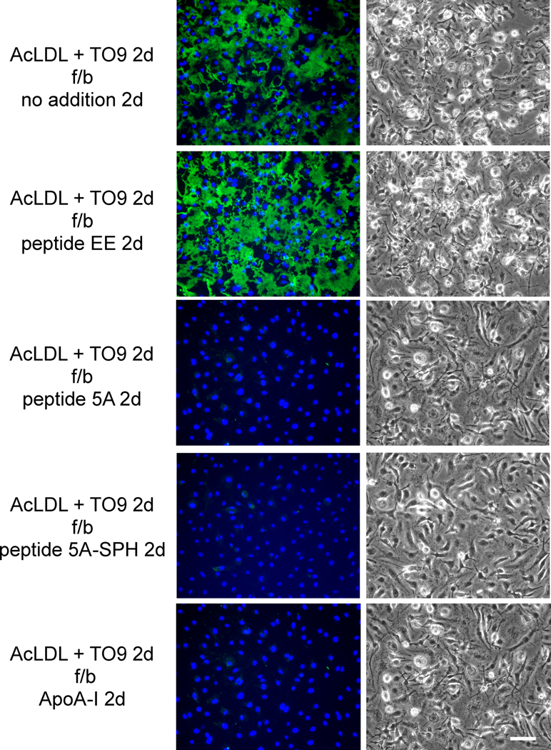
Figure 4. ApoA-I and ApoA-I mimetic peptide 5A mobilize extracellular cholesterol microdomains deposited by ABCA+/+ mouse macrophages.
One-week-old ABCA1+/+ mouse M-CSF differentiated bone marrow-derived macrophage cultures were incubated 2 days with 50 μg/ml AcLDL + 5 μm TO9 to enrich macrophages with cholesterol. Following rinsing, macrophage cultures were incubated 2 days with the indicated additions (20 μg ApoA-I, 100 μg/ml peptide, and 125 μg/ml sphingomyelin complexed with 100 μg/ml peptide 5A) without AcLDL. In the left column, cholesterol microdomains were visualized by fluorescence microscopy using anti-cholesterol microdomain Mab 58B1 (green), and nuclei were imaged with DAPI fluorescence staining (blue). In the right column, macrophages were visualized using phase-contrast microscopy. Left and right columns show corresponding microscopic fields. AcLDL, acetylated low-density lipoprotein; TO9, TO901317; f/b, followed by; 5A-SPH; 5A-sphingomyelin complex. Bar = 50 μm and applies to all.

