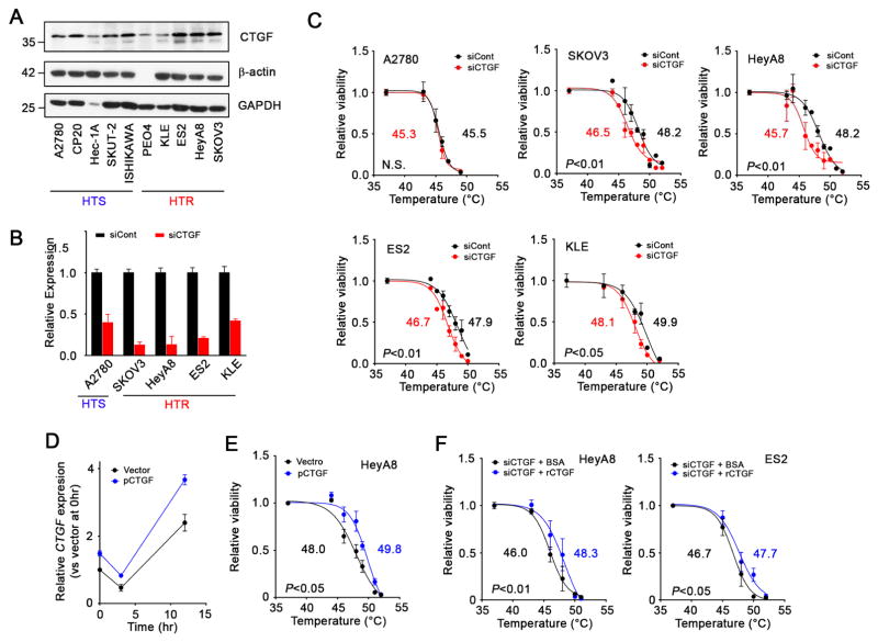
Figure 4. The effect of silencing of CTGF on sensitization of cancer cells to hyperthermia in vitro.
(A) Immunoblot of CTGF expression in tested ovarian and uterine cancer cell lines. (B) Graphs of the silencing of CTGF in tested cell lines by siRNA. Cells were transfected with siRNA at 40 nM. mRNA expression levels in the cells were evaluated 48 hr after transfection using qRT-PCR. (C) The effect of silencing of CTGF gene on sensitization of the cells to hyperthermia. Cells treated with siRNA for 48 hr were treated with hyperthermia at the indicated temperatures for 1 hr. Cells were further incubated at 37°C and their viability was determined using an MTT assay 24 hr after the initiation of hyperthermia. LT50s were calculated using a data set at least three times. (D) Time profile of CTGF mRNA expression in HeyA8 cells treated with CTGF expressing vector as determined using qRT-PCR. (E) The effect of CTGF expression on sensitization of cells to hyperthermia. HeyA8 cells that were treated with CTGF expressing vector or a control vector for 48hr were subjected to hyperthermia at the indicated temperature for 1 hr. Cell viability was determined 24 hr after the beginning of hyperthermic treatment. (F) The effect of recombinant CTGF (rCTGF) treatment on hyperthermia sensitivity. HeyA8 and ES2 cells were treated with rCTGF (200 ng/ml) 3 hr after the beginning of hyperthermic treatment. Cell viability was determined at 24 hr after the initiation of hyperthermia.
See also Figures S2 and S4

