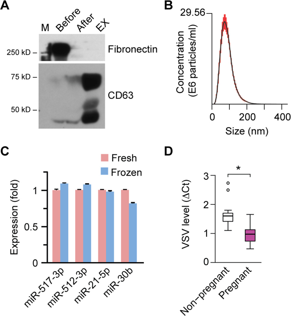
Fig 6. The effect of EXs derived from plasma of pregnant women on VSV replication in U2OS cells.
(A) A western blotting analysis depicting the removal of fibronectin, performed as a part of gelatin-agarose chromatography for purification of plasma EXs. The human plasma EXs express CD63, a canonical exosome marker. (B) NTA of purified human plasma exosomes, showing the representative distribution of exosomes in the nanometer size range (30~200 nm). (C) The expression of several representative miRNAs in fresh versus frozen plasma (n=3). None of the differences were significant. (D) The effect of plasma EXs on viral replication in U2OS cells, comparing EXs from plasma of pregnant women to EXs from plasma of non-pregnant women. Viral infection was assessed by qPCR measurement of VSV RNA levels (n=7 for each group). Linear mixed effect model was employed to evaluate the statistical difference of plasma EXs of pregnant versus non-pregnant women. *denotes p<0.05.

