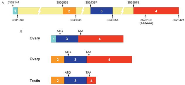
Figure 3.
(A) Putative exon–intron structure of mouse SSB-1. Published cDNA and genomic sequences of SSB-1 were compared putative exon–intron map. Nucleotide positions are as in NT_039268.4. A putative polyadenylation sequence that may account for the truncation of exon 4 in testis is indicated. (B) Structure of SSB-1 transcripts in ovary and testis as determined by RT-PCR using exon-specific primers and sequencing. Ovarian transcripts contain either exon 1 or exon 2 but not both. The coding sequence, indicated by textured bars, begins within exon 3 and ends within exon 4 and is identical in all transcripts.

