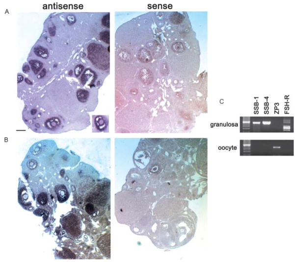
Figure 4.
(A and B) Analysis of SSB-1 (A) and SSB-4 (B) expression by in situ hybridization. Ovaries were fixed in 4% para-formaldehyde, embedded in paraffin and sectioned at 7 μm thickness. The right side shows results using sense-strand probes. Hybridization signals are concentrated over granulosa cells of primary and more advanced follicles. The inset in panel A shows an absence of signal over the oocyte. (C) Analysis of SSB-1 and SSB-4 expression by RT-PCR. Purified populations of granulosa cells and oocytes were collected and subjected to RT-PCR using primers corresponding to SSB-1, SSB-4, ZP-3 and the FSH receptor. SSB-1 and SSB-4 are expressed in granulosa cells but are not detectable in oocytes. Restriction enzyme digestion confirmed that the SSB-1 primers did not amplify SSB-4 cDNA and vice versa (not shown). The absence of ZP3 signal in granulosa cells confirms the absence of contaminating oocytes, and the absence of FSH-R signal in oocytes confirms the absence of granulosa cells.

