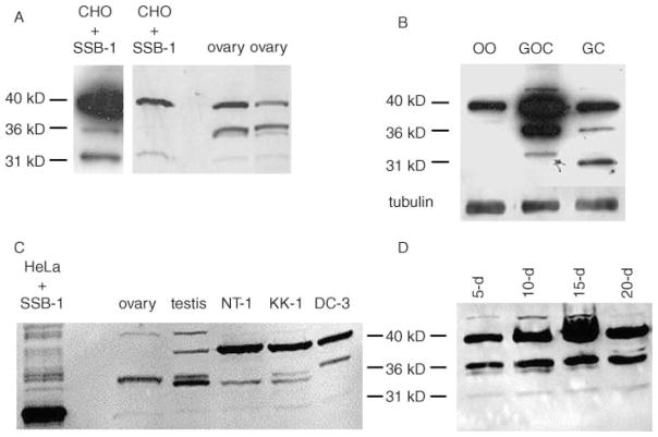
Figure 6.
Detection of SSB-1 in ovarian granulosa cells and cell lines. (A) Comparison of transfected CHO cells and whole-ovary extracts blotted using anti-SSB-1. The left panel shows a long exposure of a different CHO cell blot from that shown in the right panel. Both contain the 31, 36 and 40 kDa immunoreactive species. (B) Ovaries were digested using collagenase to yield granulosa–oocyte complexes or using collagenase and trypsin to dissociate the cell types. Oocytes (OO), complexes (GOC) or granulosa cells (GC) were collected and immunoblotted. The 31 and 36 kDa species are present in complexes and purified granulosa cells but not in purified oocytes. The increased mobility of the 31 kDa species in the granulosa cell lane was not reproducible. (C) Extracts were prepared from whole ovaries and testes and from immortalized granulosa cell lines: NT-1 and KK-1 from mouse and DC3 from rat. The 31 and 36 kDa species are present in all groups, with the latter apparently existing as a doublet. (D) Granulosa–oocyte complexes were collected from mice at different ages (shown in days), yielding populations enriched for complexes at successively more advanced stages of folliculogenesis. SSB-1 is present at all stages of follicular growth.

