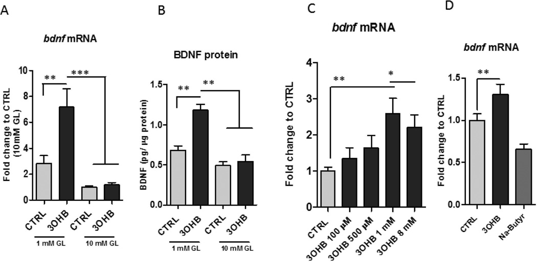
Figure 3. 3OHB treatment increases BDNF expression in cerebral cortical neurons.
Cortical neurons were incubated in medium containing either a low (1 mM) or high (10 mM) concentration of glucose (GL) and were then exposed to 8 mM 3OHB or vehicle control (CTRL). Neurons were then harvested after either 6 hours of 3OHB treatment for measurement of Bdnf mRNA levels (A) or 24 hours for measurement of BDNF protein levels (B). Values are the mean and SEM of determinations made in 5 separate experiments. **p<0.01, ***p<0.001. C. Bdnf gene expression levels were increased by 3OHB in primary cortical neurons in a concentration-dependent manner in 1 mM glucose condition. ***p<0.001 D. No induction of Bdnf gene by Na-butyrate (8 mM) was detected in 1mM glucose condition.

