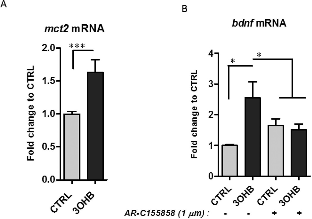
Figure 4. 3OHB treatment increases BDNF expression in cerebral cortical neurons in a MCT2-dependent manner.
A. Cortical neurons were incubated in medium containing 1 mM glucose and were then exposed to 8 mM 3OHB for 6 hours. Levels of mct2 mRNA were quantified (n =5 separate cultures). ***p<0.001. B. Cortical neurons (in medium containing 1 mM glucose) were pre-incubated with or without the MCT2 inhibitor AR-C155858 (1 µM) for 1 hour. The neurons were then treated with 8 mM 3OHB or vehicle control for 6 hours and were then processed for measurement of Bdnf mRNA levels (n = 4 separate cultures). *p<0.05.

