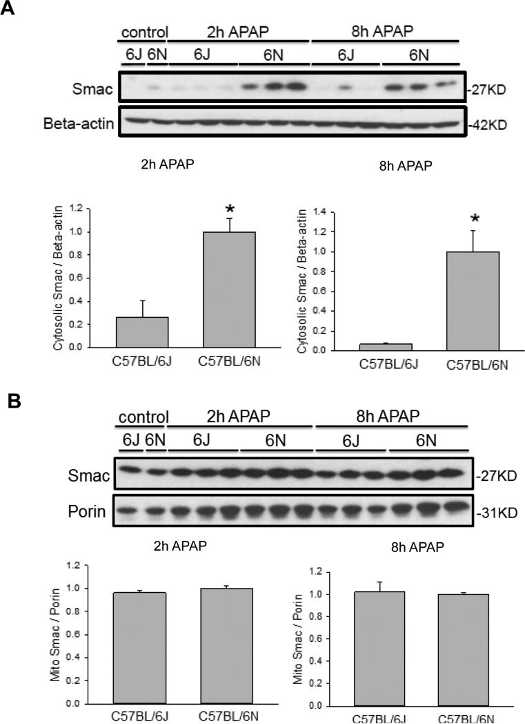
Figure 7.
Mitochondrial SMAC release after APAP treatment in C57BL/6J and C57BL/6N mice. Animals were treated with 200 mg/kg APAP or saline as control. At 2 h and 8 h after APAP, cytosolic fractions were subjected to western blotting for SMAC and ß-actin (A). Western blotting was also performed on the isolated mitochondrial fraction for SMAC and porin (B). Densitometry was performed on these blots and the cytosolic SMAC-to-beta actin and the mitochondrial SMAC-to-porin ratio was calculated for the different time points of C57BL/6J and C57BL/6N mice. The cytosolic or mitochondrial ratio for each time point for the 6N strain was set to 1 and the corresponding ratios for the 6J strain were expressed as a fraction of 1. Data represent means ± SE of n=3 animals per group. *p< 0.05 (compared with C57BL/6J mice, t=0).

