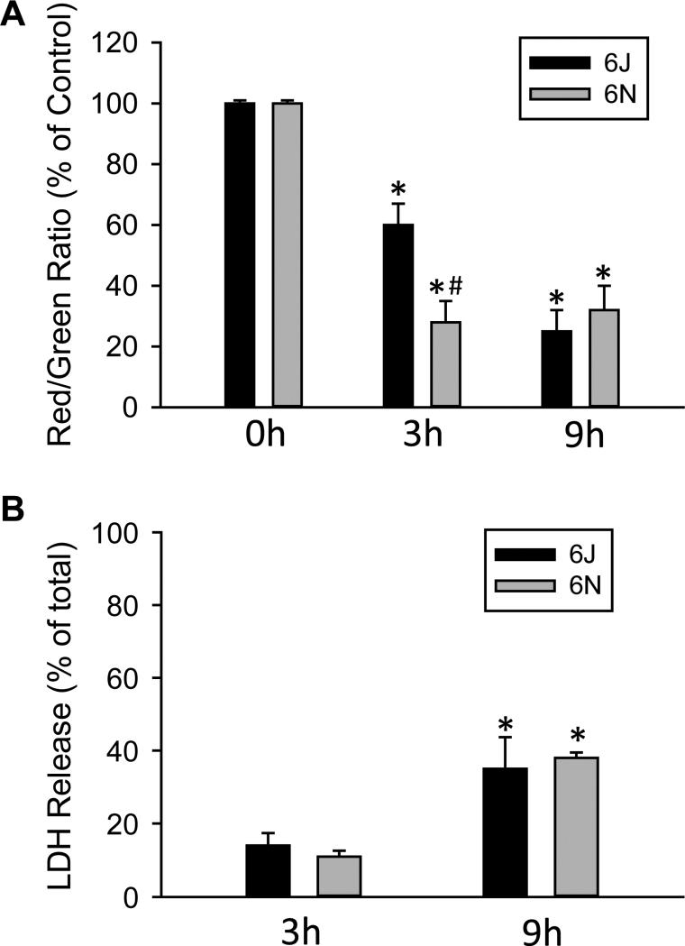
Figure 9.
Effect of APAP on mitochondrial function and viability in cells from C57BL/6J and C57BL/6N mice. Primary hepatocytes isolated from C57BL/6J and C57BL/6N mice were treated with media containing 5 mM APAP or control media. (A) Mitochondrial membrane potential, as indicated by the red/green fluorescence ratio, was determined with the JC-1 assay at 0, 3 and 9 h after 5 mM APAP. (B) Cell death, as indicated by the percentage of lactate dehydrogenase (LDH) released into the culture media, was assessed at 3 and 9 h after APAP. Data represent mean ± SE of n = 4 cell isolations. *p < 0.05 (compared to untreated cells, t=0); #p< 0.05 (compared to cells from C57BL/6J mice).

