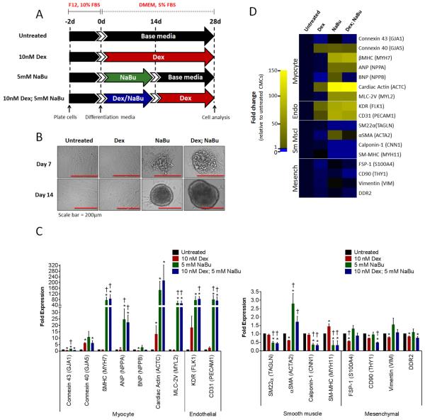
Figure 3.
NaBu-treated CMCs assume a cardiomyogenic/vasculogenic cell-like fate in vitro. (A): In vitro cardiogenic differentiation schematic and timeline. Untreated, dexamethasone (Dex), sodium butyrate (NaBu), or combination (Dex; NaBu) treated CMCs were cultured under reduced serum conditions for 28 ds to induce cardiogenic differentiation. (B): Representative phase contrast images of untreated, Dex, NaBu, or combination treated CMCs at 7 and 14 ds post induction of differentiation. (C): Real-time qPCR assays assessing the expression of various cardiovascular cell lineages (myocyte, endothelial, smooth muscle and mesenchymal/fibroblast) in differentiated CMCs. Values are mean (n=8) ± SEM. * P <0.05 (Relative to untreated) and † P <0.05 (Relative to Dex). (D): Heat map summarizing resultant qPCR-based gene expression assays. Yellow indicates an increase in fold change (>1) and blue a decrease in fold change (<1).

